
How the Test is Performed
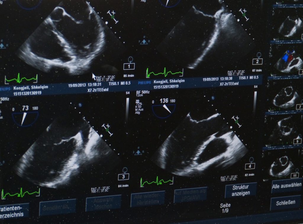
Ultrasound scanning is performed by a specifically trained Sonographer, who uses the ultrasound machine to obtain images on a screen, usually in black and white. These images are stored electronically and can be printed out for viewing, but most of the information is gained by an experienced sonographer during the actual examination.
For an ultrasound of the upper abdomen (liver, gallbladder, pancreas, spleen and kidneys) the patient is usually asked to fast for four hours before the scan. If a pelvic ultrasound is to be performed (usually to examine the uterus and ovaries in a woman) the patient needs to have a full bladder.
The patient is usually required to change into a hospital gown. A special gel is applied to the abdomen, to assist with ultrasound transmission. The gel may be cold but is otherwise harmless. Occasionally, mild pressure of the probe during the examination can be uncomfortable, if the area being examined is tender.
Get on top of your general health
Find and instantly book affordable GPs within Australia
Medical Conditions and Symptoms
An Ultrasound Scan of the Abdomen is often requested by a doctor in the setting of abdominal pain where the cause is not clear. It is the preferred test to confirm or exclude gallstones, which may cause symptoms of pain in the right upper abdomen (often going through to the back), and less commonly, jaundice (yellow discolouration of the eyes and skin), pale stools and very dark urine. Gallstones may also result in an inflammation or infection of the gallbladder, known as cholecystitis, which causes a more constant pain in the right upper abdomen and often a fever. Ultrasound is a reasonably good way to examine the kidneys for size and shape, for example in the setting of a urinary tract infection, high blood pressure (hypertension), or kidney failure. Ultrasound is also used to evaluate and monitor various forms of liver disease including cirrhosis and cancer.
Abdominal Ultrasound is frequently ordered in women with lower abdominal pain, to exclude an abnormality of the uterus or ovaries (such as ovarian cysts).
Abdominal Ultrasound is the main imaging modality in pregnancy, to assess the growth and wellbeing of the unborn baby. Ultrasound can be used to estimate the gestation of the pregnancy (how many weeks pregnant you are) and therefore the expected date of delivery – this is most accurate early in the pregnancy. A scan usually performed at 12 weeks helps to determine the risk of Down’s Syndrome. It measures the thickness of a stripe at the back of the fetal neck and this is known as “nuchal lucency”. The “Anatomy Scan”, usually done at around 18 weeks, aims to identify any structural abnormalities in the unborn fetus, and gives information about the placenta as well.
Test Results Explained
In general, a NEGATIVE scan is good news which means that the scan is normal.
An Ultrasound Scan may be POSITIVE for a specific condition, for example, gallstones, which means that an abnormality or disease has been identified.
The report and conclusion usually focus on this abnormality.
General comments about other normal organs is usually made, and sometimes limitations of the scan are described. An example of this would include appendicitis. The appendix itself is often very difficult to see on ultrasound, and the report would usually state that the appendix has not been visualised, and appendicitis is therefore not excluded.
Related Specialists
- Radiologist
- General Practitioner (GP)
- General Surgeon
- Emergency Physician
- General Physician
- Obstetrician
- Gynaecologist
- Urologist
Related Procedures
- Laparoscopy
- Endoscopic Retrograde Cholangiopancreatography (ERCP)
- Laparoscopic Surgery
- Cholecystectomy
Related Tests
- Pelvic Ultrasound Scan
- Abdominal X-Ray (AXR)
- Amylase
- Barium Swallow
- Colonoscopy
- CT Scan of the Abdomen
- Endoscopy of the Upper Gastrointestinal Tract
- Faecal Occult Blood (FOB)
- Full Blood Count (FBC)
- Gastrografin Swallow
- Hepatitis Serology (HepA, HepB, HepC)
- Lipase
- Liver Function Tests (LFT)
- Urea & Electrolytes (U&E)
- Urea Breath Test for Helicobacter pylori
- Urinalysis (UA)
- Urine HCG (Urine Pregnancy Test)
Also Known As
- Abdominal Ultrasound
- Abdo Ultrasound
- Abdominal Sonography
- Abdominal Sonogram
- Abdominal Sonar
- Abdominal US
- Abdominal U/S
- Abdominal USS
Links
A: Use HealthEngine to find and book your next GP appointment. Click on the following locations to find a GP clinic in your state or territory.
This article is for informational purposes only and should not be taken as medical advice. If in doubt, HealthEngine recommends consulting with a registered health practitioner.
All content and media on the HealthEngine Blog is created and published online for informational purposes only. It is not intended to be a substitute for professional medical advice and should not be relied on as health or personal advice. Always seek the guidance of your doctor or other qualified health professional with any questions you may have regarding your health or a medical condition. Never disregard the advice of a medical professional, or delay in seeking it because of something you have read on this Website. If you think you may have a medical emergency, call your doctor, go to the nearest hospital emergency department, or call the emergency services immediately.
Related Articles


Need a health appointment?
Find and book a doctor, dentist, physio and more on Healthengine
Find a practitioner


