
How the Test is Performed
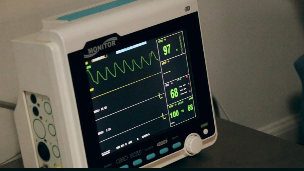
An ECG is a relatively simple test which involves sticking about ten small electrodes on the chest and attaching them to a machine at the bedside. The machine analyses the heart’s electrical signals, picked up through the electrodes on the skin surface. It is a completely painless test but obviously requires temporary exposure of the chest, to allow placement of the leads. The actual reading takes about one minute and a printout with 12 waveforms is produced either at the machine itself, or at a nearby printer. A doctor or experienced and specialised nurse reads and interprets the ECG. Sometimes they will seek additional help from a more senior doctor or a cardiologist – even at a distance via fax. Occasionally the ECG is formally reported by a cardiologist – this is more likely in the routine or non-urgent situation.
Book your health appointments online
Find and instantly book your next health appointment with Healthengine
Medical Conditions and Symptoms
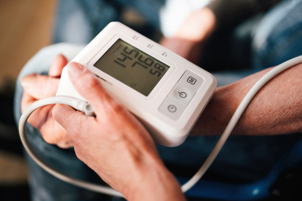
An Electrocardiogram (ECG) may be ordered as a routine test, for example before an operation to assess the electrical function of the heart, or as an urgent test in the setting of chest pain or other cardiovascular symptoms (such as shortness of breath or palpitations).
Test Results Explained
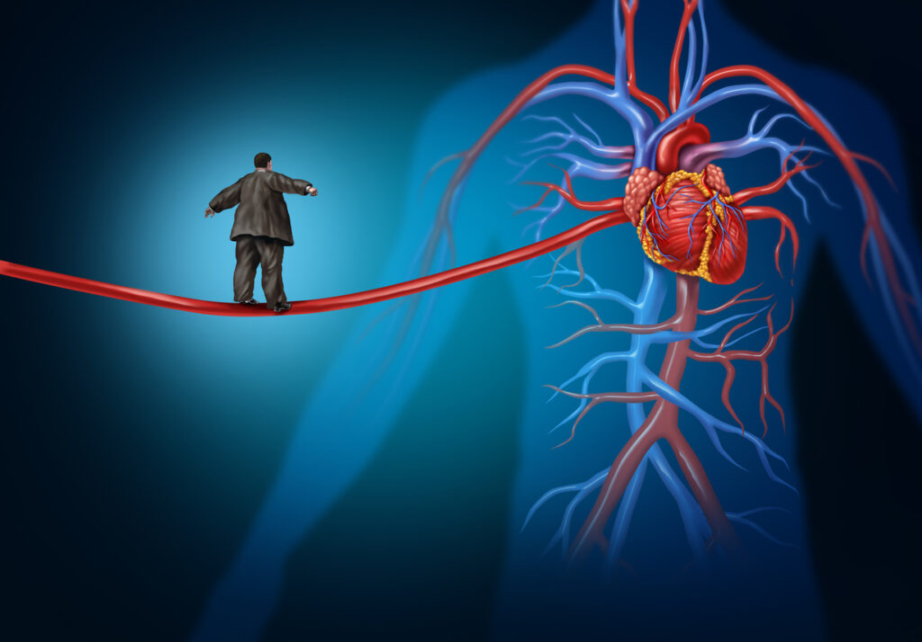
The Electrocardiogram (ECG) is a very confusing array of squiggles and waveforms, to the uninitiated. There is a large amount of information conveyed by the ECG, as each separate bump or wave can be assessed for size, shape, and distance from other bumps on the ECG. It can show abnormalities of heart rate, rhythm, axis (the relative dominance of the left or right side of the heart), and conduction of impulses through the heart. The ECG can also show the presence of damage to the heart muscle, as in a heart attack, myocarditis, or pericarditis. It is also used to diagnose angina or an acute coronary syndrome, where inadequate blood supply to the heart muscle manifests as a strain on the heart (myocardial ischaemia).
Related Specialists
- General Practitioner (GP)
- Cardiologist
- Emergency Physician
- General Physician
- Anaesthetist
- Intensivist
- Paediatrician
Related Procedures
- Pre-operative Anaesthetic assessment
- Percutaneous Coronary Intervention (PCI)(=PTCA: Percutaneous Coronary Angioplasty – with or without coronary stent placement)
- Coronary Artery Bypass Grafting (CABG)
Related Tests
- Exercise Stress Test (EST)
- Dipyridamole-Thallium Scan (Dip-Thall)
- Echocardiogram (Echo)
- Chest X-Ray (CXR)
- Troponin
- Lipid Profile (Cholesterol Test)
- CT Pulmonary Angiogram (CTPA)
- Endoscopy of the Upper Gastrointestinal Tract (Upper GI Endoscopy)
Also Known As
- EKG
- Cardiograph
Links
This article is for informational purposes only and should not be taken as medical advice. If in doubt, HealthEngine recommends consulting with a registered health practitioner.
All content and media on the HealthEngine Blog is created and published online for informational purposes only. It is not intended to be a substitute for professional medical advice and should not be relied on as health or personal advice. Always seek the guidance of your doctor or other qualified health professional with any questions you may have regarding your health or a medical condition. Never disregard the advice of a medical professional, or delay in seeking it because of something you have read on this Website. If you think you may have a medical emergency, call your doctor, go to the nearest hospital emergency department, or call the emergency services immediately.
Related Articles

Need a health appointment?
Find and book a doctor, dentist, physio and more on Healthengine
Find a practitioner