
Cervical Spine X-Ray
Last updated: 29 November 2017
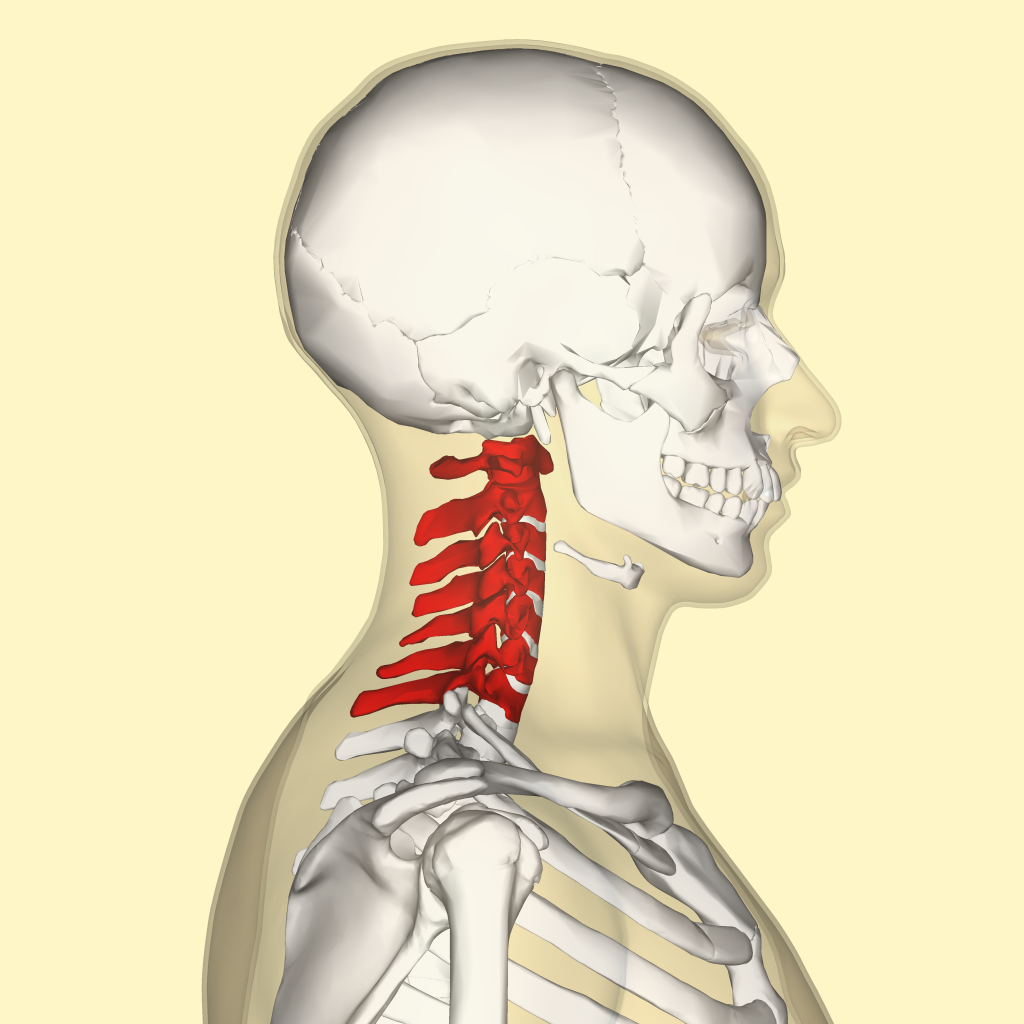
What is a cervical spine x-ray?
Cervical spine x-ray (XR cervical spine or CS spine) is performed to investigate neck pain, particularly following trauma or in cases of chronic neck pain with findings of upper limb weakness, numbness or tingling.
How is a cervical spine x-ray performed?
Cervical spine x-ray is performed by a radiographer in an X-Ray room. The standard three views taken are the:
- AP (anteroposterior view, which looks at the spine from the front)
- Lateral (which looks at the spine from the side)
- Peg view (this looks at the upper part of the cervical spine and requires the patient to open the mouth wide)
X-Rays are taken with the patient’s head in full flexion (leaning as far forward as possible). The patient will be asked to bend the head forward as far as possible, and to extend the neck backwards as far as possible.
Find and instantly book affordable GPs within AustraliaGet on top of your general health
When would you need a cervical spine x-ray?
A cervical spine x-ray is useful in identifying cervical vertebrae fractures (bone breaks), vertebral malalignment, dislocation and degenerative spine disease.
Is a spinal x-ray done for various sections of the spine?
A doctor or technical can performing imaging for different parts of the spine for a closer look at things. The spine is made up of smaller bones called vertebrae. The imaging professional may do x-rays for cervical spine (neck), thoracic spine (chest or trunk area), lumbar spine (lower back), or the sacral area (base of the spine), as well as the Coccyx (tailbone).
What problems will a spinal xray reveal?
A spinal x-ray can reveal things such as spinal fractures, disk problems, infections, tumors, abnormal curvature of the spine, Scoliosis, arthritis and pretty much anything that may be impacting the spine negatively, including congenital issues that a person may be born with.
Test results, explained
In cases of trauma, the cervical spine x-ray is usually interpreted immediately by an emergency doctor such as an Emergency Physician, an Orthopaedic Surgeon, or a General Surgeon involved in the care of the patient. Sometimes, a Radiologist may also be requested to interpret the images.
C-Spine X-Rays taken in the non-urgent setting are reported by a Radiologist, and the report is usually sent to the doctor who ordered the test. The Radiologist will usually comment on the alignment of the vertebrae and the presence or absence of any ‘wear and tear’ changes, usually related to normal aging and known as degenerative changes or osteoarthritis.
Related specialists
- General Practitioner (GP)
- Radiologist
- Emergency physician
- Orthopaedic surgeon
- Neurosurgeon
- Neurologist
Related procedures
- Pre-operative Anaesthetic assessment
Related tests
Also known as
- XR cervical spine
- Davis series cervical spine
- C spine X-Ray
- C spine series
- AP cervical spine
- Lateral cervical spine
- Flexion/extension views
- 5-series cervical spine
- 7-series cervical spine
Links
A: Use HealthEngine to find and book your next Radiologist appointment. Click on the following locations to find a Radiologist clinic in your state or territory.
This article is for informational purposes only and should not be taken as medical advice. If in doubt, HealthEngine recommends consulting with a registered health practitioner.
All content and media on the HealthEngine Blog is created and published online for informational purposes only. It is not intended to be a substitute for professional medical advice and should not be relied on as health or personal advice. Always seek the guidance of your doctor or other qualified health professional with any questions you may have regarding your health or a medical condition. Never disregard the advice of a medical professional, or delay in seeking it because of something you have read on this Website. If you think you may have a medical emergency, call your doctor, go to the nearest hospital emergency department, or call the emergency services immediately.
Related Articles


Need a health appointment?
Find and book a doctor, dentist, physio and more on Healthengine
Find a practitioner